Our Facilities

24 X 7 Emergency Care Facility.

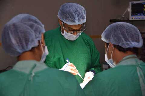
We provides emergent, urgent, and sub-urgent care to adult, pediatric, and obstratics patients. The 24 Hour Emergency Department is equipped to handle any medical emergencies around the clock every day. Our team of multi-disciplinary paramedics, nurses and doctors are at hand to ensure your condition is properly assessed and managed. Our specialists and surgeons are also on call 24 hours to help attend to your medical condition.

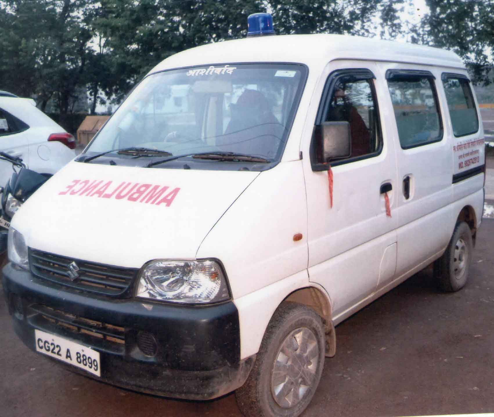
24 X 7 Ambulance Facility.

The nearest ambulance available in your locality will be rushed to your place, wherever you are, to bring your patient directly to Chanda Devi Tiwari hospital in the shortest possible time. And while the patient is on the way the Emergency Team at CDTH will keep the ICU Bed, O T, etc, ready so that no time is lost when the patient reaches hospital . No other hospital in Baloda bazar offers 24-hrs ambulance pick-up service. Not even during daytime .

Express Service For Emergency Patient.

Express service not only saves your time it also makes you free from the rush and allows you to directly consult with your respected physician to get better diagnosis and allows you to get a royality messages about the on calls Doctors visits at our centre for your better health .

Medicine

24x7 Pharmacy Services/ Medical store

The mission of the pharmacy is to provide medications, other health care products, relevant information, professional services and to help people and society to make the best use of them. Every activity in the pharmacy is done with certain system and confidence, in order to give the right touch of professionalism and care.

We are best in delivering medicines, services and care of the patient.

Parking Facility Available

The mission of Parking Services is to provide courteous, safe, and efficient parking services for the campus community and public.Parking service is free for all 2 as well as 4 wheelers 24 x 7 .
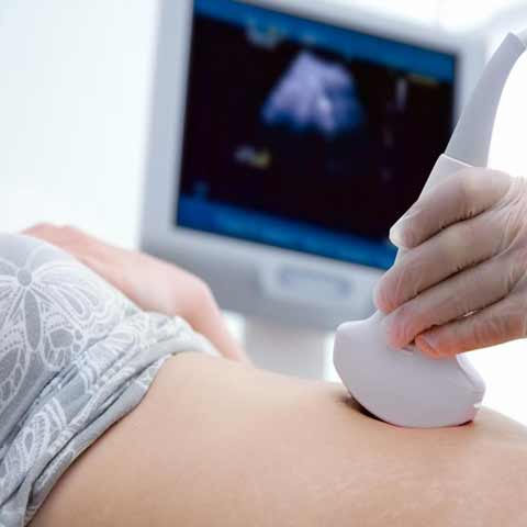
Sonography

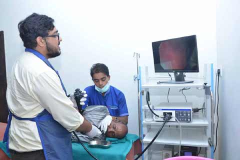
Ultrasound imaging uses sound waves to produce pictures of the inside of the body. It is used to help diagnose the causes of pain, swelling and infection in the body’s internal organs and to examine a baby in pregnant women and the brain and hips in infants. It’s also used to help guide biopsies, diagnose heart conditions, and assess damage after a heart attack. Ultrasound is safe, noninvasive, and does not use ionizing radiation.

Ultrasound is safe and painless, and produces pictures of the inside of the body using sound waves. Ultrasound imaging, also called ultrasound scanning or sonography, involves the use of a small transducer (probe) and ultrasound gel placed directly on the skin. High-frequency sound waves are transmitted from the probe through the gel into the body. The transducer collects the sounds that bounce back and a computer then uses those sound waves to create an image. Ultrasound examinations do not use ionizing radiation (as used in x-rays), thus there is no radiation exposure to the patient. Because ultrasound images are captured in real-time, they can show the structure and movement of the body's internal organs, as well as blood flowing through blood vessels.
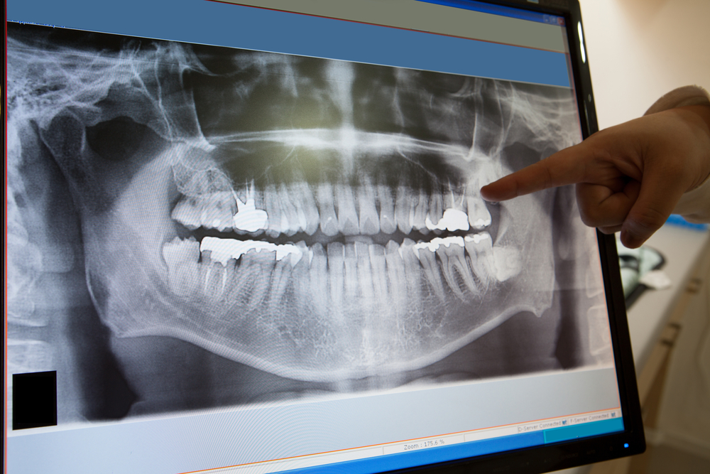
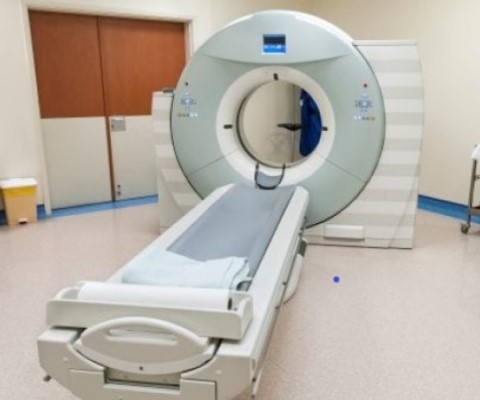
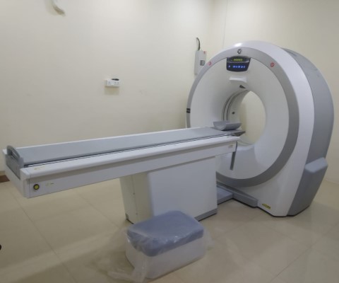
Digital X-Ray

X-rays are a form of electromagnetic radiation, as are radio waves, infrared radiation, visible light, ultraviolet radiation and microwaves. One of the most common and beneficial uses of X-rays is for medical imaging. X-rays are also used in treating cancer and in exploring the cosmos.

Electromagnetic radiation is transmitted in waves or particles at different wavelengths and frequencies. This broad range of wavelengths is known as the electromagnetic spectrum. The EM spectrum is generally divided into seven regions in order of decreasing wavelength and increasing energy and frequency. The common designations are: radio waves, microwaves, infrared (IR), visible light, ultraviolet (UV), X-rays and gamma-rays.
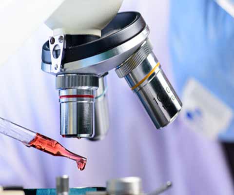
Pathology Lab

Scientific Pathology offer a wide range of quality assured clinical and pathology investigation services using the latest technology, state of art equipments and testing facilities manned by experienced / trained doctors and technicians.

CT- Scan

CT-Scan Facility

A computed tomography (CT or CAT) scan allows doctors to see inside your body. It uses a combination of X-rays and a computer to create pictures of your organs, bones, and other tissues. It shows more detail than a regular X-ray.

You can get a CT scan on any part of your body. The procedure doesn't take very long, and it's painless.

They use a narrow X-ray beam that circles around one part of your body. This provides a series of images from many different angles. A computer uses this information to create a cross-sectional picture. Like one piece in a loaf of bread, this two-dimensional or three-dimensional (2D OR 3D) scan shows a “slice” of the inside of your body.

This process is repeated to produce a number of slices. The computer stacks these scans one on top of the other to create a detailed image of your organs, bones, or blood vessels. For example, a surgeon may use this type of scan to look at all sides of a tumor to prepare for an operation.

Smart Card Facility

Social Security and healthcare assurance for all has been the motto of Government of India, and it has taken various steps in this regard. One of the most important policy milestones is the Unorganized Workers Social Security Act (2008) enacted by the Central Government to provide for the social security and welfare of the unorganized workers. This act recommends that the Central Government provide social security schemes to mitigate risks due to disability, health shocks, maternity and old age which all unorganized workers get exposed to and are likely to suffer from.

Janani Suraksha Yojana

Janani Suraksha Yojana (JSY) is a safe motherhood intervention under the National Rural Health Mission (NRHM) being implemented with the objective of reducing maternal and neo-natal mortality by promoting institutional delivery among the poor pregnant women. The Yojana, launched on 12th April 2005, by the Hon’ble Prime Minister, is being implemented in all states and UTs with special focus on low performing states. JSY is a 100 % centrally sponsored scheme and it integrates cash assistance with delivery and post-delivery care.

Laparoscopy & Minimal Access Surgery

Dr. Nitin Tiwari specializes in laparoscopic surgery and hysteroscopy from the onset of his career. He performs surgery with over 5 years of expertise at CDTH hospital which have the most modern technology

Laparoscopy has been used in gynecology as a diagnostic method since 1970. Laparoscopy is an operation performed in the abdomen or pelvis through small incisions (usually 0.5–1.5 cm) with the aid of a camera. It can either be used to inspect and diagnose a condition or to perform surgery.

Hystroscopy

An endoscopy is a procedure in which your doctor uses specialized instruments to view and operate on the internal organs and vessels of your body. It allows surgeons to view problems within your body without making large incisions.

A surgeon inserts an endoscope through a small cut, or an opening in the body such as the mouth. An endoscope is a flexible tube with an attached camera that allows your doctor to see. Your doctor can use forceps (tongs) and scissors on the endoscope to operate or remove tissue for biopsy.

Endoscopy

Non-robotic minimally invasive surgery (endoscopic)

Non-robotic minimally invasive surgery is also known as endoscopic surgery. You also may be familiar with terms like laparoscopic surgery, thoracoscopic surgery, or “keyhole” surgery. These are minimally invasive procedures that utilize an endoscope to reach internal organs through very small incisions. During endoscopic surgery the surgeon inserts a thin, flexible tube with a video camera through a small incision or a natural orifice like the mouth or nostrils. The tube has a channel to utilize tiny surgical instruments, which the surgeon uses while viewing the organs on a computer monitor.

ECG

An electrocardiogram is a picture of the electrical conduction of the heart. By examining changes from normal on the ECG, clinicians can identify a myriad of cardiac disease processes. There are two ways to learn ECG interpretation — pattern recognition (the most common) and understanding the exact electrical vectors recorded by an ECG as they relate to cardiac electrophysiology — and most people learn a combination of both. This tutorial pairs the approaches. Basing ECG interpretation on pattern recognition alone is often not sufficient.

Respiratory and Chest

Respiratory and Chest

In the respiratory examination, the patient is asked to sit upright on an examination table, with arms at the side. Adequate lighting is ensured and the patient is asked to expose his or her chest.

Later in the examination, when the back is examined, the patient is usually asked to move the arms forward so that the scapulae are not in the way of examining the upper lung fields. These fields are intended to correlate with the lung lobes and are thus tested on the anterior and posterior chest walls (the front and back of the patient's thorax).

The respiratory exam has conventionally been split into different stages:

CT- Scan

MULTISLICE CT SCAN FACILITY AVAILABLE 24 X 7 HOURS

Trauma Facility in Chanda Devi Triwari Hospital

TRAUMA FACILITY AVAILABLE 24 X 7 HOURS

The Best Medical Service in BalodaBazar

We Provide the Best Medical Service

Call Us: 07727-222125 For Emergency